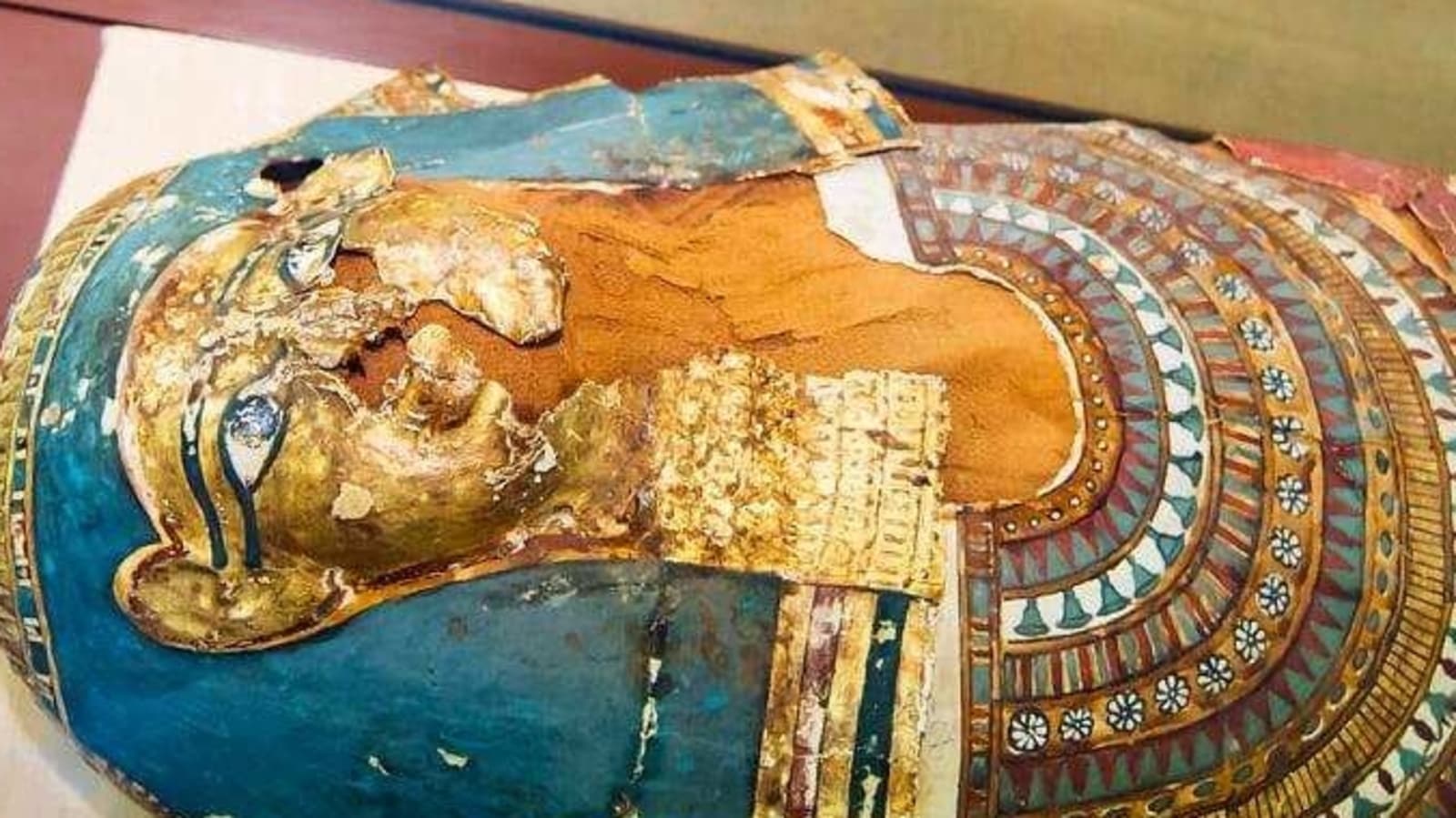
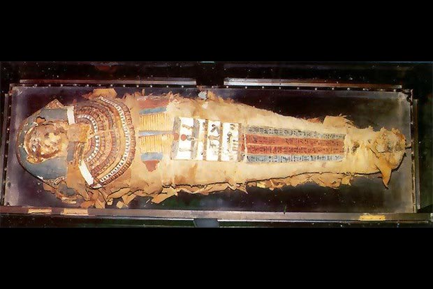
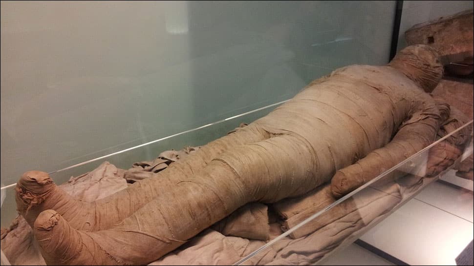
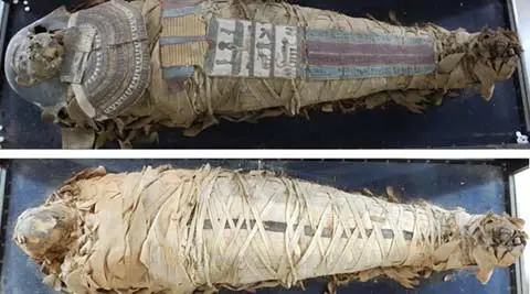
With the aim of stopping the decay of an over 2,000 year-old Egyptian mummy preserved at the Telangana State Museum, the Department of Archaeology and Museums has begun its preventive conservation using unique and advanced scientific techniques.
This technique, involving CT scan and X-ray examination, is being used on a decaying mummy in India for the first time, according to officials.

The mummy was obtained by the sixth Nizam, Mir Mehboob Ali Khan, in 1920. His son and the last Nizam, Mir Osman Ali Khan, donated it to the museum, where it has been kept since 1930.
The mummy at this museum is one of only six Egyptian mummies preserved in museums across the country. It was previously believed to be that of a 16-18 year old girl from the Ptolemaic period (300 BC to 100 BC).
“The scan revealed that the mummy was actually that of a 25 year old woman about 136 cm in height,” Secretary (Youth Advancement, Tourism and Culture Department) B Venkatesham told reporters last evening.

The conservation efforts undertaken for the mummy represent a significant step forward in preservation techniques, particularly in India. Vinod Daniel, the Heritage Conservation Adviser to the project, highlighted the pioneering nature of the conservation methods, which can serve as an example for similar projects both nationally and internationally. By placing the mummy in an oxygen-free encasement, further decay can be prevented, and the original materials have been carefully preserved as part of the conservation process.
The mummy’s painted cartonnage pieces were meticulously restored to their proper position, and the showcase was sealed to ensure protection from external elements. The damage to the mummy, caused by various factors including heat, light, temperature, humidity, insects, and oxygen, will be minimized within the oxygen-free case, where bacteria and insects cannot survive, and humidity levels remain stable.
The use of advanced techniques such as CT scans and X-rays has provided valuable insights into the mummy’s condition, allowing for informed decisions regarding its conservation and display. The microscopic photograph and CT scan report will be made available to the public for the first time, contributing to broader understanding and appreciation of the mummy’s significance.
Despite some damage to the ribs, spine, ankle, and the presence of a metallic amulet, the majority of the mummy’s bones, skull, and teeth remain intact. The next phase of treatment will focus on remedial conservation and potential restoration of the cartonnage, paving the way for its eventual display in a special showcase. Overall, this project serves as a comprehensive case study for the conservation of mummies worldwide, showcasing the intersection of scientific advancements and cultural heritage preservation.


